
Introduction
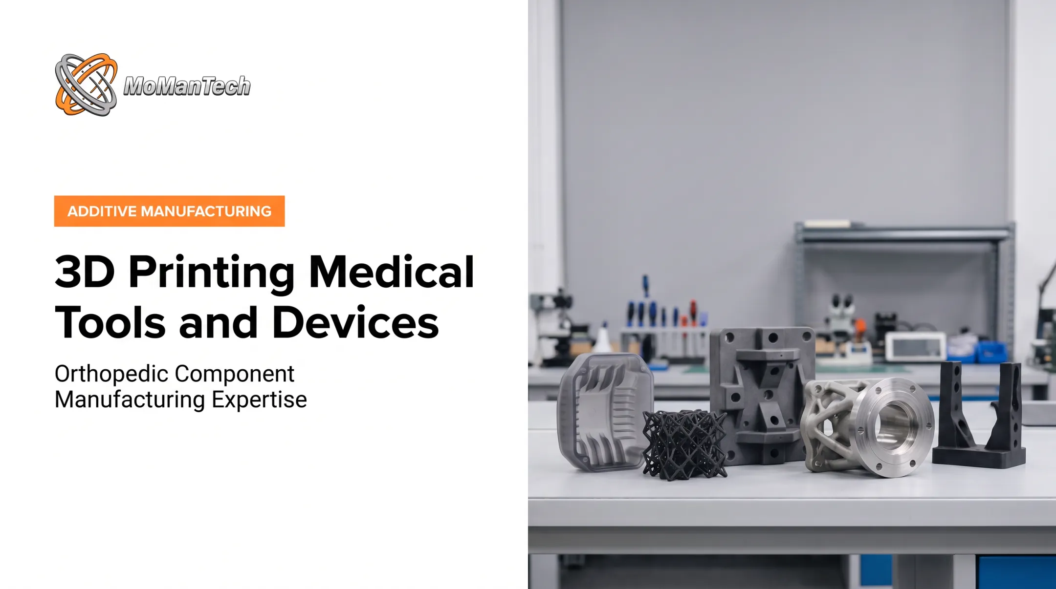
Standard implants work well for standard anatomy. But revision hips with large bone loss, tumor resections, and irregular defects often outrun what off-the-shelf sizes can handle. Additive manufacturing (AM) builds geometry that conventional cutting tools can't reach, from porous bone-contact surfaces to patient-matched shapes.
Yet the technology isn't a shortcut. A 2024 acetabular review found no significant pooled improvement in Harris Hip Score for 3D-printed cups across three nonrandomized comparisons.
This article covers current U.S. applications, materials and design, the manufacturing workflow, regulatory and quality considerations, and how OEMs can evaluate an additive or hybrid production partner.
Key Takeaways
- Additive manufacturing enables patient-matched implants and porous structures that are hard to machine.
- It pays off when anatomy, geometry, or low volumes justify extra validation.
- Materials, post-processing, inspection, and regulatory evidence matter as much as the print.
- Precision machining often finishes printed parts, so the two methods complement each other.
What Is 3D Printing in Orthopedics?
Additive manufacturing builds a part layer by layer from a digital model. The FDA describes the process as sequentially building and joining layers. Machining does the opposite: it removes material from stock.
The Typical Digital Workflow
For patient-matched devices, the path usually runs:
- Patient imaging (CT or MRI) and image processing
- CAD design within a defined design envelope
- Engineering review and build preparation
- Printing
- Post-processing (heat treatment, support removal, finishing)
- Final testing and inspection
Standard-size AM implants skip the imaging steps but follow the rest.
Implants Versus Everything Else
Categories often blur, so keep them distinct:
- Implanted devices: cages, acetabular cups, augments, and patient-specific reconstructions
- External prosthetic limbs and orthotics: worn outside the body, with a different evidence base
- Surgical guides: instruments used during surgery, covered by FDA's May 2026 final guidance on patient-matched guides
- Bioprinted tissue: FDA's 2017 AM guidance excludes biological, cellular, and tissue-based products, and these remain largely experimental
Where Are 3D-Printed Orthopedic Implants Used?
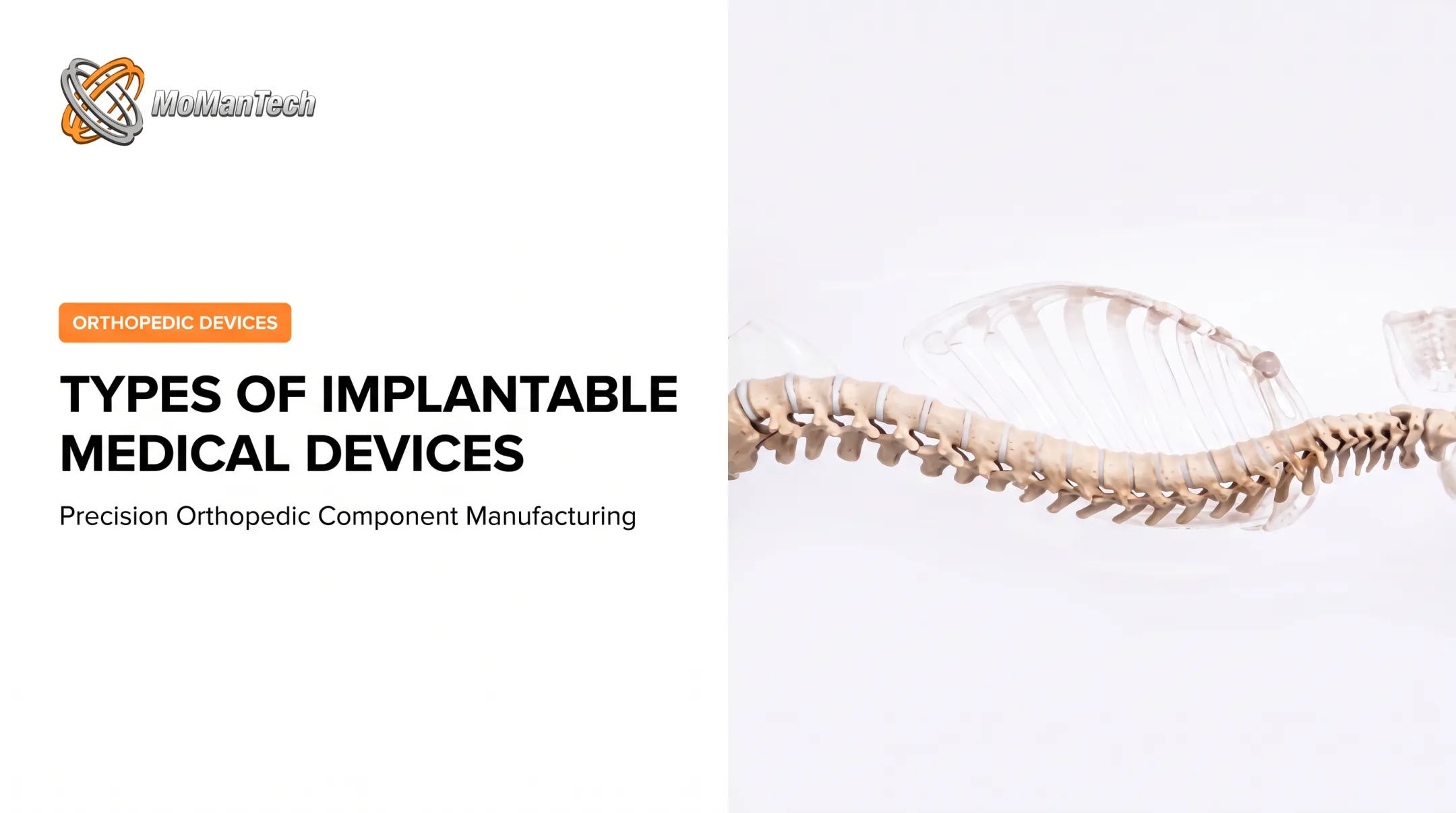
FDA records document printed spinal, hip, knee, pelvic, and craniofacial implants. Each clearance is product-specific.
| Anatomy | Example | FDA status |
|---|---|---|
| Lumbar interbody | Stryker Tritanium PL | 510(k) K181014, 2018 |
| Cervical interbody | Curiteva Porous PEEK cage | 510(k) K213030, 2023 |
| Acetabular shell | Smith+Nephew REDAPT | 510(k) K182109, 2018 |
| Acetabular augment | REDAPT Augments | 510(k) K171073, 2017 |
| Knee tibial baseplate | Stryker Triathlon Tritanium | 510(k) K123486, 2013 |
| Patient-specific pelvis | Onkos My3D | 510(k) K223348, 2023 |
| Cranial | OsteoFab (PEKK) | 510(k) K121818, 2013 |
| Facial | OsteoFab (PEKK) | 510(k) K161052, 2016 |
Spine and Joint Reconstruction
Spinal cages use controlled porosity to combine solid and porous structures in one body. In joints, printed acetabular shells and augments address bone deficiencies where a cup alone isn't enough.
Trauma, Extremity, and Complex Defects
Patient-specific designs suit irregular bone loss, tumor resection, and difficult foot and ankle anatomy. Cranial and facial defects fit scan-to-design workflows well because anatomical fit, fixation, and imaging compatibility drive the design.
Case study: patient-specific talus. For talar avascular necrosis, identifiable CT landmarks allow a patient-specific design. The implant is additively manufactured from cobalt-chromium and then polished. Per FDA's 2021 summary, the device received Humanitarian Device Exemption approval (H200001), not 510(k) clearance.
Single-center data covered 31 patients and 32 cases. Mean pain scores fell from 6.9 cm to 4.1 cm on a visual analog scale, with 3 reoperations. That supports a probable-benefit finding, not superiority over conventional reconstruction.

Established Versus Emerging
Status by category:
- Metal and polymer implants: cleared or approved, and established in use
- Porous ceramic scaffolds: mostly still research
- Bioprinted bone: not publicly confirmed as a U.S.-cleared product
The AuriNovo ear-cartilage trial (NCT04399239) was terminated for a company decision, not safety, and it concerned cartilage, not bone.
Benefits and Tradeoffs of 3D-Printed Orthopedic Implants
Fit, Porosity, and Evidence
Patient-matched design helps surgeons plan for unusual anatomy. Customization alone doesn't guarantee better outcomes, as the acetabular data above show.
Porosity affects ingrowth. A rabbit study tested 300, 600, and 900 micrometer pores at 65% target porosity, The 600 micrometer group showed greater early fixation at two weeks. That's animal data, not a universal human specification.
Porous designs might also reduce stress shielding. But a 2023 systematic review found 45 of 46 studies were computational or in vitro, with only one clinical study. Strength and fatigue limits still apply.
AM Versus Conventional Machining
| Factor | Additive | Machining |
|---|---|---|
| Design freedom | Lattices, internal geometry | Limited by tool access |
| Tooling | Less dedicated tooling | Fixtures and programming |
| Surface finish | Usually needs post-processing | Strong for smooth surfaces |
| Tolerance | Must be demonstrated per device | Strong for tight critical dimensions |
| Volume | Often suits complex, low-volume work | Efficient for repeat batches |
| Waste | Builds material; powder and supports need control | Removes material from stock |
Neither method is always faster or cheaper. Geometry, quantity, and finishing decide.
Materials and Manufacturing Workflow
Selecting Materials
Material suitability depends on strength, fatigue life, corrosion resistance, imaging compatibility, biocompatibility, and implant location.
- Titanium alloys: ASTM F2924 covers Ti-6Al-4V by powder bed fusion; F3001 covers the extra-low-interstitial grade
- Cobalt-chromium: ASTM F3213 covers additive Co-28Cr-6Mo; ASTM F75 covers cast CoCr, so the two aren't interchangeable
- Stainless steel: ASTM F3184 covers UNS S31603 but explicitly does not meet surgical implant-grade S31673
- PEEK/PEKK: used in porous cervical cages and in PEKK cranial and facial devices
- Ceramics and bioactive materials: hydroxyapatite, TCP, and bioactive glass remain mostly research scaffolds
Designing for Additive Manufacturing
Designers balance several additive-specific factors:
- Load case and fixation method
- Pore geometry and wall thickness
- Build orientation and support structures
- Thermal distortion and finishing allowances
Priorities shift by implant type. A spinal cage emphasizes porosity and endplate contact. A joint augment emphasizes fixation surfaces. A talus spacer needs smooth articulating surfaces after polishing.

Printing and Post-Processing
FDA's 2017 guidance expects documentation of:
- Starting powder identity, supplier, reuse, and virgin-to-used ratios
- Build orientation, location, and machine parameters
- Heat treatment or hot isostatic pressing, which can reduce porosity and improve fatigue life
- Support removal, finishing, and final machining
Tortuous pores complicate cleaning and sterilization, so FDA recommends testing worst-case geometry on final-finished devices. Processes whose output can't be fully verified by later inspection need validation.
Inspection, Testing, and Traceability
Verification may include dimensional inspection, surface characterization, CT or micro-CT, and mechanical testing. ASTM F2077 covers intervertebral fusion devices and F1717 covers spinal constructs. Material certificates, lot traceability, and first-article records round out the file.
Where Precision Machining Fits
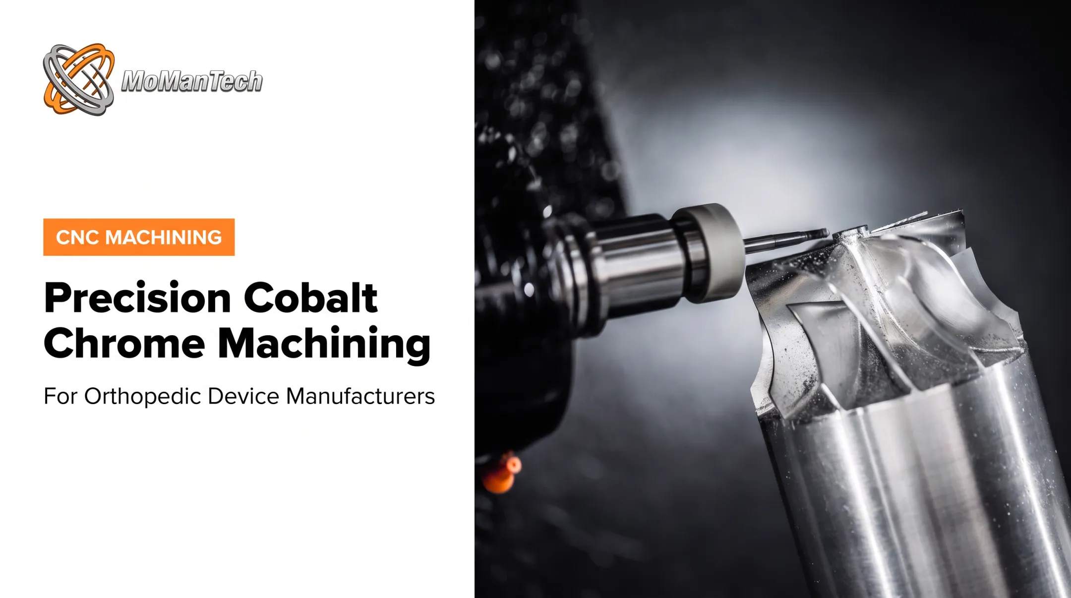
ASTM F2924 explicitly lists machining, grinding, EDM, and polishing to reach critical dimensions and surface finish. A hybrid workflow prints complex bulk or porous geometry, then machines holes, threads, and mating interfaces.
MoManTech, an ISO 13485:2016-certified contract manufacturer focused exclusively on orthopedic devices, covers the machining side of that workflow. It offers precision CNC, wire EDM, design-for-manufacturability support, documented inspection, and prototype-to-production runs from 1 to 100+ units.
Quality, Regulation, and Commercialization Considerations
U.S. Regulatory Pathway
Device classification, intended use, and risk determine the FDA pathway. Additive manufacturing is a production method, not a regulatory pathway on its own. Options include 510(k), De Novo, PMA, and HDE, as the talus case shows.
Patient matching doesn't automatically qualify for the custom-device exemption, which is narrow and capped at 5 units per year of a device type.
Quality Management and Validation
The Quality Management System Regulation took effect February 2, 2026, incorporating ISO 13485:2016. Your QMS should cover:
- Design controls and supplier qualification
- Equipment qualification and build-process validation
- Software change control
- Nonconformance handling and corrective actions
Biological and Mechanical Safety
FDA's 2023 ISO 10993-1 guidance calls for risk-based biological evaluation of the final-finished device, including sterilization effects. Build residual powder, cleaning, corrosion, fatigue, and wear testing into that plan.
Clinical Evidence and Responsible Claims
Keep evidence levels separate: bench tests, animal studies, early clinical series, and cleared devices are not interchangeable. Avoid promising faster recovery or fewer complications without supporting data.

Evaluating a Manufacturing Partner
Ask whether the partner offers:
- Orthopedic experience in your device category
- Design-for-manufacturability input before design freeze
- Material and process knowledge, including post-processing
- Prototype and first-article support
- Documented inspection and traceability
- Responsive communication
- Capacity to repeat orders with consistent processes
Frequently Asked Questions
What is 3D printing in orthopedics?
It's layer-by-layer manufacturing of orthopedic devices from digital models. Imaging and CAD can produce patient-specific designs.
Are 3D printed prosthetics better?
Not necessarily. A 2025 review of 11 studies and 177 participants couldn't draw firm conclusions, and breakage appeared in four studies. External prostheses also differ from implanted devices.
Are 3D printed orthotics good?
They can offer customized fit and pressure offloading. Effectiveness still depends on assessment, design, material, and professional fitting.
Is it possible to 3D print bones?
Printed bone-like scaffolds exist, but living, functional printed bone does not. Bone bioprinting remains research.
Has 3D bioprinting been successful?
Early trials exist, such as the terminated AuriNovo ear study. Printing living tissue differs entirely from printing metal or polymer implants.
How much do 3D printed prosthetics cost?
Costs vary with device type, materials, customization, fitting, and maintenance. No reliable U.S. range is established, and prosthetic prices shouldn't be applied to implants.